

Is Endoplasmic Reticulum Found In Plant Or Animal Cells
| Jail cell biology | |
|---|---|
| Fauna cell diagram | |
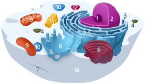 Components of a typical animal cell:
|

Micrograph of rough endoplasmic reticulum network around the nucleus (shown in the lower right-hand area of the film). Dark pocket-sized circles in the network are mitochondria.
The endoplasmic reticulum (ER) is, in essence, the transportation system of the eukaryotic cell, and has many other important functions such as protein folding. It is a type of organelle made up of ii subunits – rough endoplasmic reticulum (RER), and smooth endoplasmic reticulum (SER). The endoplasmic reticulum is found in near eukaryotic cells and forms an interconnected network of flattened, membrane-enclosed sacs known as cisternae (in the RER), and tubular structures in the SER. The membranes of the ER are continuous with the outer nuclear membrane. The endoplasmic reticulum is non found in red blood cells, or spermatozoa.
The two types of ER share many of the same proteins and engage in certain mutual activities such every bit the synthesis of certain lipids and cholesterol. Unlike types of cells contain different ratios of the 2 types of ER depending on the activities of the cell. RER is establish mainly toward the nucleus of jail cell and SER towards the jail cell membrane or plasma membrane of prison cell.
The outer (cytosolic) face up of the RER is studded with ribosomes that are the sites of protein synthesis. The RER is especially prominent in cells such every bit hepatocytes. The SER lacks ribosomes and functions in lipid synthesis but not metabolism, the production of steroid hormones, and detoxification.[ane] The SER is peculiarly abundant in mammalian liver and gonad cells.
The ER was observed with low-cal microscope past Garnier in 1897, who coined the term ergastoplasm.[2] [iii] With electron microscopy, the lacy membranes of the endoplasmic reticulum were first seen in 1945 by Keith R. Porter, Albert Claude, and Ernest F. Fullam.[iv] Later, the discussion reticulum, which means "network", was applied by Porter in 1953 to depict this cloth of membranes.[5]
Structure [edit]
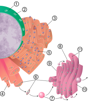
i Nucleus 2 Nuclear pore three Rough endoplasmic reticulum (RER) iv Smoothen endoplasmic reticulum (SER) 5 Ribosome on the rough ER 6 Proteins that are transported vii Transport vesicle 8 Golgi apparatus 9 Cis confront of the Golgi apparatus 10 Trans face of the Golgi appliance 11 Cisternae of the Golgi apparatus

3D rendering of endoplasmic reticulum
The full general construction of the endoplasmic reticulum is a network of membranes called cisternae. These sac-like structures are held together past the cytoskeleton. The phospholipid membrane encloses the cisternal space (or lumen), which is continuous with the perinuclear space but split up from the cytosol. The functions of the endoplasmic reticulum can be summarized equally the synthesis and consign of proteins and membrane lipids, just varies between ER and cell type and cell office. The quantity of both rough and smooth endoplasmic reticulum in a cell can slowly interchange from i type to the other, depending on the changing metabolic activities of the cell. Transformation can include embedding of new proteins in membrane likewise as structural changes. Changes in poly peptide content may occur without noticeable structural changes.[six] [7] [ commendation needed ]
Rough endoplasmic reticulum [edit]
![]()
A 2-minute blitheness showing how a protein destined for the secretory pathway is synthesized into the rough endoplasmic reticulum, which appears at the upper right approximately halfway through the blitheness.
The surface of the rough endoplasmic reticulum (often abbreviated RER or rough ER; besides called granular endoplasmic reticulum) is studded with poly peptide-manufacturing ribosomes giving it a "crude" appearance (hence its name).[8] The binding site of the ribosome on the rough endoplasmic reticulum is the translocon.[9] However, the ribosomes are non a stable part of this organelle's structure as they are constantly being bound and released from the membrane. A ribosome only binds to the RER one time a specific protein-nucleic acrid complex forms in the cytosol. This special circuitous forms when a free ribosome begins translating the mRNA of a protein destined for the secretory pathway.[10] The start 5–30 amino acids polymerized encode a signal peptide, a molecular bulletin that is recognized and jump by a signal recognition particle (SRP). Translation pauses and the ribosome circuitous binds to the RER translocon where translation continues with the nascent (new) poly peptide forming into the RER lumen and/or membrane. The protein is processed in the ER lumen by an enzyme (a point peptidase), which removes the signal peptide. Ribosomes at this point may be released dorsum into the cytosol; however, non-translating ribosomes are also known to stay associated with translocons.[11]
The membrane of the rough endoplasmic reticulum forms large double-membrane sheets that are located almost, and continuous with, the outer layer of the nuclear envelope.[12] The double membrane sheets are stacked and continued through several right- or left-handed helical ramps, the "Terasaki ramps", giving rise to a construction resembling a multi-story motorcar park.[13] [fourteen] Although there is no continuous membrane between the endoplasmic reticulum and the Golgi apparatus, membrane-bound transport vesicles shuttle proteins between these two compartments.[15] Vesicles are surrounded by coating proteins chosen COPI and COPII. COPII targets vesicles to the Golgi apparatus and COPI marks them to be brought back to the crude endoplasmic reticulum. The rough endoplasmic reticulum works in concert with the Golgi complex to target new proteins to their proper destinations. The second method of send out of the endoplasmic reticulum involves areas called membrane contact sites, where the membranes of the endoplasmic reticulum and other organelles are held closely together, allowing the transfer of lipids and other small molecules.[sixteen] [17]
The rough endoplasmic reticulum is key in multiple functions:
- Manufacture of lysosomal enzymes with a mannose-vi-phosphate marker added in the cis-Golgi network.[ citation needed ]
- Manufacture of secreted proteins, either secreted constitutively with no tag or secreted in a regulatory manner involving clathrin and paired basic amino acids in the signal peptide.
- Integral membrane proteins that stay embedded in the membrane as vesicles exit and demark to new membranes. Rab proteins are primal in targeting the membrane; SNAP and SNARE proteins are key in the fusion upshot.
- Initial glycosylation equally assembly continues. This is N-linked (O-linking occurs in the Golgi).
- Due north-linked glycosylation: If the poly peptide is properly folded, oligosaccharyltransferase recognizes the AA sequence NXS or NXT (with the Due south/T remainder phosphorylated) and adds a 14-sugar backbone (2-N-acetylglucosamine, ix-branching mannose, and 3-glucose at the stop) to the side-chain nitrogen of Asn.
Shine endoplasmic reticulum [edit]

Electron micrograph showing polish ER (pointer) in mouse tissue, at 110,510× magnification.
In almost cells the smooth endoplasmic reticulum (abbreviated SER) is scarce. Instead there are areas where the ER is partly smooth and partly rough, this surface area is called the transitional ER. The transitional ER gets its proper noun because it contains ER exit sites. These are areas where the transport vesicles that contain lipids and proteins made in the ER, disassemble from the ER and beginning moving to the Golgi appliance. Specialized cells can take a lot of smooth endoplasmic reticulum and in these cells the smooth ER has many functions.[6] It synthesizes lipids, phospholipids,[xviii] [19] [20] and steroids. Cells which secrete these products, such every bit those in the testes, ovaries, and sebaceous glands have an affluence of polish endoplasmic reticulum.[21] It also carries out the metabolism of carbohydrates, detoxification of natural metabolism products and of alcohol and drugs, attachment of receptors on cell membrane proteins, and steroid metabolism.[22] In muscle cells, it regulates calcium ion concentration. Smooth endoplasmic reticulum is plant in a variety of cell types (both animate being and plant), and it serves different functions in each. The smooth endoplasmic reticulum also contains the enzyme glucose-vi-phosphatase, which converts glucose-6-phosphate to glucose, a footstep in gluconeogenesis. It is connected to the nuclear envelope and consists of tubules that are located near the jail cell periphery. These tubes sometimes branch forming a network that is reticular in appearance.[12] In some cells, in that location are dilated areas similar the sacs of rough endoplasmic reticulum. The network of smoothen endoplasmic reticulum allows for an increased surface surface area to be devoted to the action or storage of central enzymes and the products of these enzymes.
Sarcoplasmic reticulum [edit]

The sarcoplasmic reticulum (SR), from the Greek σάρξ sarx ("flesh"), is smooth ER found in musculus cells. The only structural difference between this organelle and the shine endoplasmic reticulum is the medley of proteins they have, both bound to their membranes and globe-trotting within the confines of their lumens. This fundamental difference is indicative of their functions: The endoplasmic reticulum synthesizes molecules, while the sarcoplasmic reticulum stores calcium ions and pumps them out into the sarcoplasm when the muscle fiber is stimulated.[23] [24] Later their release from the sarcoplasmic reticulum, calcium ions interact with contractile proteins that utilize ATP to shorten the musculus cobweb. The sarcoplasmic reticulum plays a major role in excitation-contraction coupling.[25]
Functions [edit]
The endoplasmic reticulum serves many full general functions, including the folding of poly peptide molecules in sacs called cisternae and the transport of synthesized proteins in vesicles to the Golgi apparatus. Rough endoplasmic reticulum is besides involved in poly peptide synthesis. Correct folding of newly made proteins is made possible by several endoplasmic reticulum chaperone proteins, including poly peptide disulfide isomerase (PDI), ERp29, the Hsp70 family member BiP/Grp78, calnexin, calreticulin, and the peptidylprolyl isomerase family. Just properly folded proteins are transported from the crude ER to the Golgi apparatus – unfolded proteins cause an unfolded poly peptide response every bit a stress response in the ER. Disturbances in redox regulation, calcium regulation, glucose deprivation, and viral infection[26] or the over-expression of proteins[27] can atomic number 82 to endoplasmic reticulum stress response (ER stress), a state in which the folding of proteins slows, leading to an increase in unfolded proteins. This stress is emerging as a potential crusade of damage in hypoxia/ischemia, insulin resistance, and other disorders.[28]
Protein send [edit]
Secretory proteins, mostly glycoproteins, are moved beyond the endoplasmic reticulum membrane. Proteins that are transported by the endoplasmic reticulum throughout the jail cell are marked with an accost tag called a point sequence. The Northward-terminus (1 end) of a polypeptide chain (i.east., a protein) contains a few amino acids that work as an address tag, which are removed when the polypeptide reaches its destination. Nascent peptides reach the ER via the translocon, a membrane-embedded multiprotein circuitous. Proteins that are destined for places exterior the endoplasmic reticulum are packed into ship vesicles and moved along the cytoskeleton toward their destination. In homo fibroblasts, the ER is e'er co-distributed with microtubules and the depolymerisation of the latter cause its co-aggregation with mitochondria, which are also associated with the ER.[29]
The endoplasmic reticulum is also part of a poly peptide sorting pathway. It is, in essence, the transportation system of the eukaryotic prison cell. The bulk of its resident proteins are retained within it through a retention motif. This motif is equanimous of four amino acids at the end of the poly peptide sequence. The most common retention sequences are KDEL for lumen located proteins and KKXX for transmembrane protein.[30] However, variations of KDEL and KKXX practise occur, and other sequences can as well give ascent to endoplasmic reticulum retention. Information technology is not known whether such variation tin lead to sub-ER localizations. There are three KDEL (i, 2 and three) receptors in mammalian cells, and they have a very high degree of sequence identity. The functional differences betwixt these receptors remain to exist established.[31]
Bioenergetics regulation of ER ATP supply by a CaATiER machinery [edit]

Ca2+-antagonized send into the endoplasmic reticulum (CaATiER) model
The endoplasmic reticulum does not harbor an ATP-regeneration mechanism, and therefore requires ATP import from mitochondria. The imported ATP is vital for the ER to carry out its house keeping cellular functions, such as for protein folding and trafficking.[32]
The ER ATP transporter, SLC35B1/AXER, was recently cloned and characterized,[33] and the mitochondria supply ATP to the ER through a Catwo+-antagonized transport into the ER (CaATiER) mechanism.[34] The CaATiER mechanism shows sensitivity to cytosolic Ca2+ ranging from high nM to low μM range, with the Catwo+-sensing element yet to exist identified and validated.
Clinical significance [edit]
Increased and supraphysiological ER stress in pancreatic β cells disrupts normal insulin secretion, leading to hyperinsulinemia[35] and consequently peripheral insulin resistance associated with obesity in humans.[36]
Abnormalities in XBP1 lead to a heightened endoplasmic reticulum stress response and later on causes a higher susceptibility for inflammatory processes that may even contribute to Alzheimer'south disease.[37] In the colon, XBP1 anomalies have been linked to the inflammatory bowel diseases including Crohn's disease.[38]
The unfolded protein response (UPR) is a cellular stress response related to the endoplasmic reticulum.[39] The UPR is activated in response to an accumulation of unfolded or misfolded proteins in the lumen of the endoplasmic reticulum. The UPR functions to restore normal function of the cell past halting protein translation, degrading misfolded proteins, and activating the signaling pathways that lead to increasing the production of molecular chaperones involved in protein folding. Sustained overactivation of the UPR has been implicated in prion diseases too as several other neurodegenerative diseases and the inhibition of the UPR could become a treatment for those diseases.[40]
References [edit]
- ^ "Endoplasmic Reticulum (Rough and Smooth)". British Gild of Prison cell Biology. Archived from the original on 24 November 2015. Retrieved 21 November 2015.
- ^ Garnier, C. 1897. Les filaments basaux des cellules glandulaires. Note préliminaire. Bibliographie anatomique, 5, 278–289.
- ^ Buvat R (1963). "Electron microscopy of plant protoplasm". International Review of Cytology. xiv: 41–55. doi:10.1016/S0074-7696(08)60021-2. ISBN9780123643148. PMID 14283576.
- ^ Porter KR, Claude A, Fullam EF (March 1945). "A study of tissue culture cells past electron microscopy: methods and preliminary observations". The Journal of Experimental Medicine. 81 (3): 233–46. doi:10.1084/jem.81.3.233. PMC2135493. PMID 19871454.
- ^ PORTER KR (May 1953). "Observations on a submicroscopic basophilic component of cytoplasm". The Journal of Experimental Medicine. 97 (v): 727–50. doi:10.1084/jem.97.5.727. PMC2136295. PMID 13052830.
- ^ a b Alberts B, Johnson A, Lewis J, Raff M, Roberts Thou, Walter P (2002). Molecular biological science of the prison cell (4th ed.). New York: Garland Science. ISBN978-0-8153-3218-three. Archived from the original on 2017-10-03.
- ^ Cooper GM (2000). The cell: a molecular arroyo (2nd ed.). Washington (DC): ASM Printing. ISBN978-0-87893-106-4.
- ^ "reticulum". The Complimentary Dictionary.
- ^ Görlich D, Prehn S, Hartmann Eastward, Kalies KU, Rapoport TA (October 1992). "A mammalian homolog of SEC61p and SECYp is associated with ribosomes and nascent polypeptides during translocation". Jail cell. 71 (3): 489–503. doi:ten.1016/0092-8674(92)90517-Chiliad. PMID 1423609. S2CID 19078317.
- ^ Lodish H, et al. (2003). Molecular Cell Biology (fifth ed.). West. H. Freeman. pp. 659–666. ISBN978-0-7167-4366-eight.
- ^ Seiser RM, Nicchitta CV (October 2000). "The fate of membrane-bound ribosomes following the termination of protein synthesis". The Journal of Biological Chemical science. 275 (43): 33820–7. doi:x.1074/jbc.M004462200. PMID 10931837.
- ^ a b Shibata Y, Voeltz GK, Rapoport TA (Baronial 2006). "Rough sheets and smooth tubules". Cell. 126 (3): 435–9. doi:ten.1016/j.cell.2006.07.019. PMID 16901774. S2CID 16107069.
- ^ Terasaki Thou, Shemesh T, Kasthuri N, Klemm RW, Schalek R, Hayworth KJ, Hand AR, Yankova 1000, Huber G, Lichtman JW, Rapoport TA, Kozlov MM (July 2013). "Stacked endoplasmic reticulum sheets are connected by helicoidal membrane motifs". Cell. 154 (2): 285–96. doi:x.1016/j.cell.2013.06.031. PMC3767119. PMID 23870120.
- ^ Guven J, Huber One thousand, Valencia DM (October 2014). "Terasaki screw ramps in the rough endoplasmic reticulum". Concrete Review Letters. 113 (xviii): 188101. Bibcode:2014PhRvL.113r8101G. doi:10.1103/PhysRevLett.113.188101. PMID 25396396.
- ^ Endoplasmic reticulum. (due north.d.). McGraw-Hill Encyclopedia of Science and Technology. Retrieved September xiii, 2006, from Answers.com Web site: "Answers - the Near Trusted Identify for Answering Life's Questions". Answers.com. Archived from the original on 2006-11-16. Retrieved 2006-09-xiii .
- ^ Levine T (September 2004). "Short-range intracellular trafficking of small molecules across endoplasmic reticulum junctions". Trends in Jail cell Biology. 14 (9): 483–90. doi:10.1016/j.tcb.2004.07.017. PMID 15350976.
- ^ Levine T, Loewen C (August 2006). "Inter-organelle membrane contact sites: through a drinking glass, darkly". Current Opinion in Prison cell Biology. 18 (4): 371–eight. doi:10.1016/j.ceb.2006.06.011. PMID 16806880.
- ^ Prinz, William A.; Choudhary, Vineet; Liu, Li-Ka; Lahiri, Sujoy; Kannan, Muthukumar (2017-03-01). "Phosphatidylserine synthesis at membrane contact sites promotes its transport out of the ER". Periodical of Lipid Research. 58 (3): 553–562. doi:10.1194/jlr.M072959. ISSN 0022-2275. PMC5335585. PMID 28119445.
- ^ Kannan, Muthukumar; Riekhof, Wayne R.; Voelker, Dennis R. (2015). "Send of Phosphatidylserine from the Endoplasmic Reticulum to the Site of Phosphatidylserine Decarboxylase2 in Yeast". Traffic. 16 (two): 123–134. doi:10.1111/tra.12236. ISSN 1600-0854. PMID 25355612. S2CID 34302.
- ^ Friedman, Jonathan R.; Kannan, Muthukumar; Toulmay, Alexandre; Jan, Calvin H.; Weissman, Jonathan S.; Prinz, William A.; Nunnari, Jodi (2018-01-22). "Lipid Homeostasis Is Maintained by Dual Targeting of the Mitochondrial PE Biosynthesis Enzyme to the ER". Developmental Cell. 44 (2): 261–270.e6. doi:10.1016/j.devcel.2017.11.023. ISSN 1534-5807. PMC5975648. PMID 29290583.
- ^ "Functions of Shine ER". University of Minnesota Duluth.
- ^ Maxfield FR, Wüstner D (Oct 2002). "Intracellular cholesterol transport". The Journal of Clinical Investigation. 110 (7): 891–8. doi:10.1172/JCI16500. PMC151159. PMID 12370264.
- ^ Toyoshima C, Nakasako M, Nomura H, Ogawa H (June 2000). "Crystal structure of the calcium pump of sarcoplasmic reticulum at ii.6 A resolution". Nature. 405 (6787): 647–55. Bibcode:2000Natur.405..647T. doi:ten.1038/35015017. PMID 10864315. S2CID 4316039.
- ^ Goodman SR (2007-11-26). Medical Cell Biology (3rd ed.). Bookish Press. p. 69. ISBN9780080919317.
- ^ Martini F, Nath J, Bartholomew E (2014). Fundamentals of Anatomy and Physiology (10th ed.). ISBN978-0321909077.
- ^ Xu C, Bailly-Maitre B, Reed JC (October 2005). "Endoplasmic reticulum stress: cell life and expiry decisions". The Journal of Clinical Investigation. 115 (10): 2656–64. doi:10.1172/JCI26373. PMC1236697. PMID 16200199.
- ^ Kober L, Zehe C, Bode J (October 2012). "Evolution of a novel ER stress based selection system for the isolation of highly productive clones". Biotechnology and Bioengineering. 109 (ten): 2599–611. doi:ten.1002/chip.24527. PMID 22510960. S2CID 25858120.
- ^ Ozcan U, Cao Q, Yilmaz E, Lee AH, Iwakoshi NN, Ozdelen Eastward, Tuncman Thousand, Görgün C, Glimcher LH, Hotamisligil GS (Oct 2004). "Endoplasmic reticulum stress links obesity, insulin activity, and type two diabetes". Scientific discipline. 306 (5695): 457–61. Bibcode:2004Sci...306..457O. doi:x.1126/scientific discipline.1103160. PMID 15486293. S2CID 22517395.
- ^ Soltys BJ, Gupta RS (1992). "Interrelationships of endoplasmic reticulum, mitochondria, intermediate filaments, and microtubules--a quadruple fluorescence labeling written report". Biochemistry and Cell Biological science. 70 (10–11): 1174–86. doi:x.1139/o92-163. PMID 1363623.
- ^ Stornaiuolo M, Lotti LV, Borgese N, Torrisi MR, Mottola G, Martire G, Bonatti S (March 2003). "KDEL and KKXX retrieval signals appended to the same reporter protein make up one's mind different trafficking betwixt endoplasmic reticulum, intermediate compartment, and Golgi complex". Molecular Biological science of the Cell. 14 (3): 889–902. doi:x.1091/mbc.E02-08-0468. PMC151567. PMID 12631711.
- ^ Raykhel I, Alanen H, Salo Yard, Jurvansuu J, Nguyen VD, Latva-Ranta Chiliad, Ruddock Fifty (Dec 2007). "A molecular specificity code for the three mammalian KDEL receptors". The Journal of Cell Biology. 179 (vi): 1193–204. doi:x.1083/jcb.200705180. PMC2140024. PMID 18086916.
- ^ Clairmont, CA; De Maio, A; Hirschberg, CB (25 February 1992). "Translocation of ATP into the lumen of rough endoplasmic reticulum-derived vesicles and its binding to luminal proteins including BiP (GRP 78) and GRP 94". The Journal of Biological Chemical science. 267 (6): 3983–90. doi:10.1016/S0021-9258(19)50622-6. PMID 1740446.
- ^ Klein, Marie-Christine; Zimmermann, Katharina; Schorr, Stefan; Landini, Martina; Klemens, Patrick A. Westward.; Altensell, Jacqueline; Jung, Martin; Krause, Elmar; Nguyen, Duy; Helms, Volkhard; Rettig, Jens; Fecher-Trost, Claudia; Cavalié, Adolfo; Hoth, Markus; Bogeski, Ivan; Neuhaus, H. Ekkehard; Zimmermann, Richard; Lang, Sven; Haferkamp, Ilka (28 August 2018). "AXER is an ATP/ADP exchanger in the membrane of the endoplasmic reticulum". Nature Communications. 9 (1): 3489. Bibcode:2018NatCo...9.3489K. doi:10.1038/s41467-018-06003-9. PMC6113206. PMID 30154480.
- ^ Yong, Jing; Bischof, Helmut; Burgstaller, Sandra; Siirin, Marina; Tater, Anne; Malli, Roland; Kaufman, Randal J (ix September 2019). "Mitochondria supply ATP to the ER through a machinery antagonized by cytosolic Ca2+". eLife. 8. doi:x.7554/eLife.49682. PMC6763289. PMID 31498082.
- ^ Yong, Jing; Johnson, James D.; Arvan, Peter; Han, Jaeseok; Kaufman, Randal J. (August 2021). "Therapeutic opportunities for pancreatic β-jail cell ER stress in diabetes mellitus". Nature Reviews. Endocrinology. 17 (viii): 455–467. doi:10.1038/s41574-021-00510-4. ISSN 1759-5037. PMID 34163039.
- ^ van Vliet, Stephan; Koh, Han-Chow E.; Patterson, Bruce W.; Yoshino, Mihoko; LaForest, Richard; Gropler, Robert J.; Klein, Samuel; Mittendorfer, Bettina (October 2020). "Obesity Is Associated With Increased Basal and Postprandial β-Cell Insulin Secretion Fifty-fifty in the Absence of Insulin Resistance". Diabetes. 69 (10): 2112–2119. doi:10.2337/db20-0377. ISSN 1939-327X. PMC7506835. PMID 32651241.
- ^ Casas-Tinto S, Zhang Y, Sanchez-Garcia J, Gomez-Velazquez Grand, Rincon-Limas DE, Fernandez-Funez P (June 2011). "The ER stress factor XBP1s prevents amyloid-beta neurotoxicity". Human Molecular Genetics. 20 (11): 2144–threescore. doi:10.1093/hmg/ddr100. PMC3090193. PMID 21389082.
- ^ Kaser A, Lee AH, Franke A, Glickman JN, Zeissig S, Tilg H, Nieuwenhuis EE, Higgins DE, Schreiber S, Glimcher LH, Blumberg RS (September 2008). "XBP1 links ER stress to abdominal inflammation and confers genetic adventure for human being inflammatory bowel illness". Cell. 134 (5): 743–56. doi:ten.1016/j.cell.2008.07.021. PMC2586148. PMID 18775308.
- ^ Walter, Peter. "Peter Walter's Curt Talk: Unfolding the UPR". iBiology.
- ^ Moreno JA, Halliday M, Molloy C, Radford H, Verity N, Axten JM, Ortori CA, Willis AE, Fischer PM, Barrett DA, Mallucci GR (October 2013). "Oral treatment targeting the unfolded protein response prevents neurodegeneration and clinical disease in prion-infected mice". Science Translational Medicine. 5 (206): 206ra138. doi:10.1126/scitranslmed.3006767. PMID 24107777. S2CID 25570626.
External links [edit]
- Endoplasmic Reticulum
- Lipid and protein limerick of Endoplasmic reticulum in OPM database
- Animations of the various cell functions referenced hither Archived 2008-04-22 at the Wayback Automobile
Source: https://en.wikipedia.org/wiki/Endoplasmic_reticulum
Posted by: wallaceborceir.blogspot.com
0 Response to "Is Endoplasmic Reticulum Found In Plant Or Animal Cells"
Post a Comment